
Microscope-Assisted Root Canal Treatment
Learn how microscope-assisted root canal treatment uses enhanced magnification and illumination to visualise complex root canal anatomy during treatment.
Quick Links
At a Glance:
|
What it is |
Root canal treatment performed with a dental operating microscope for enhanced magnification and illumination |
|
How it helps |
Provides a detailed view of the tooth and root canal system during treatment |
|
When it is used |
Complex root anatomy, narrow or calcified canals, missed canals, retreatment and certain treatment complications |
|
What it can help identify |
Additional canals, anatomical variations, cracks, obstructions and areas that can be difficult to see without magnification |
|
Can it be used for retreatment? |
Yes. Magnification can be particularly useful when examining and treating previously root canal-treated teeth |
|
Treatment goal |
To clean, disinfect and seal the root canal system while preserving the natural tooth |
Root canal systems can be small and complex, with narrow canals, additional branches and anatomical variations that can be difficult to see during treatment.
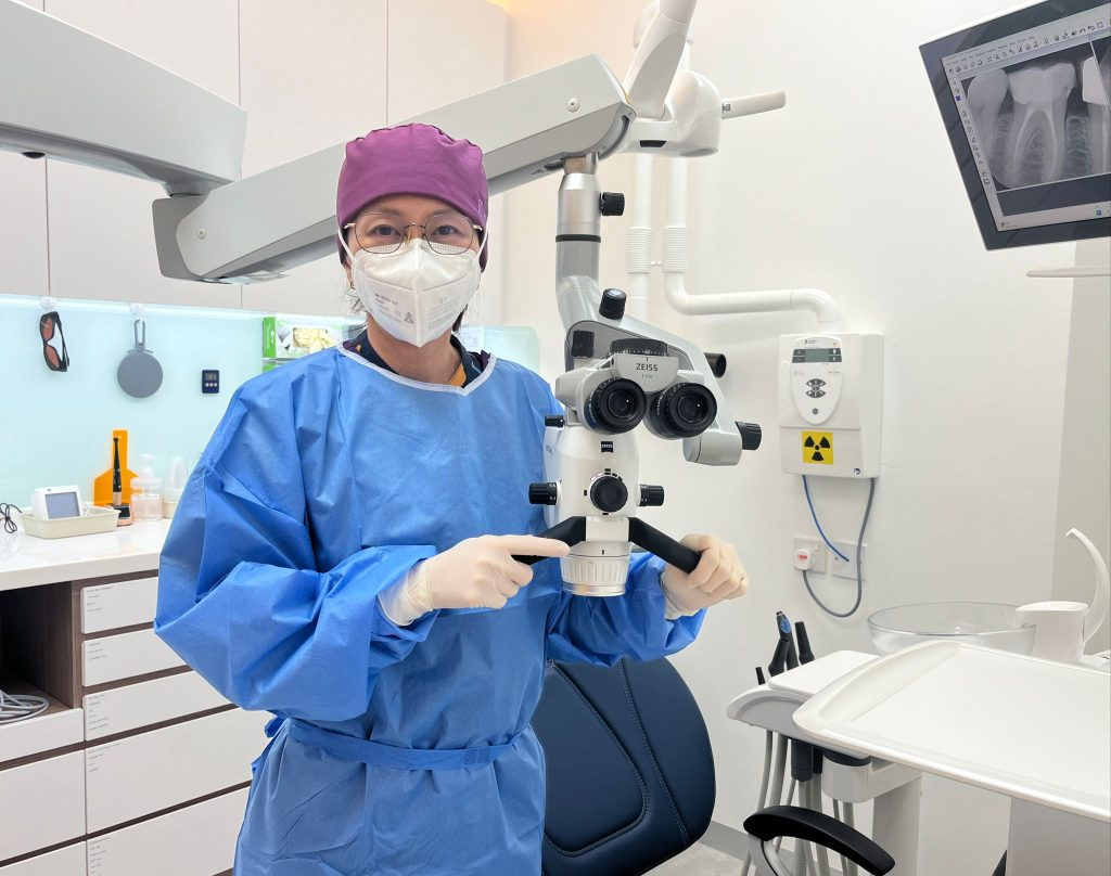
Microscope-assisted root canal treatment uses a dental operating microscope to provide enhanced magnification and illumination of the tooth and root canal system. This provides greater visualisation during the procedure and can be particularly useful when treating teeth with complex anatomy or more challenging cases.
When is microscope-assisted root canal treatment recommended?
A dental operating microscope can be used during both initial root canal treatment and retreatment. It can be particularly beneficial when the tooth has complex anatomy or when greater visualisation is needed to identify and manage specific treatment challenges.
|
When it is recommended |
How the microscope helps |
|
Complex root canal anatomy |
Provides greater visualisation of additional canals, branching and anatomical variations that can make treatment more challenging. |
|
Narrow or calcified canals |
Helps the dentist locate and access small or partially calcified canal openings that can be difficult to see. |
|
Missed or additional canals |
Can help identify canals that were not located or treated during previous root canal treatment. |
|
Root canal retreatment |
Provides a clearer view when navigating existing filling materials, restorations and changes from previous treatment. |
|
Previous treatment complications |
Can assist in identifying and managing certain complications, such as broken instruments or obstructions within the canal. |
|
Suspected cracks or fractures |
Enhanced magnification can help identify some cracks or structural defects within the visible portions of the tooth. |
What are the benefits of microscope-assisted root canal treatment?
The enhanced magnification and illumination provided by a dental operating microscope can support greater visibility and precision throughout root canal treatment.
Benefits can include:
- Enhanced visualisation — Fine details within the tooth and root canal system can be seen more clearly.
- Greater treatment precision — Magnification allows the dentist to work within a small treatment area with greater accuracy.
- Improved detection of additional canals — Small or hidden canal openings that can otherwise be difficult to identify can be more readily visualised.
- Preservation of healthy tooth structure — Greater visibility can help the dentist access and treat the root canal system more precisely while limiting unnecessary removal of surrounding tooth structure.
- Better management of complex cases — Enhanced visibility can assist when navigating calcified canals, unusual anatomy, previously treated teeth and certain treatment complications.
How is microscope-assisted root canal treatment performed?
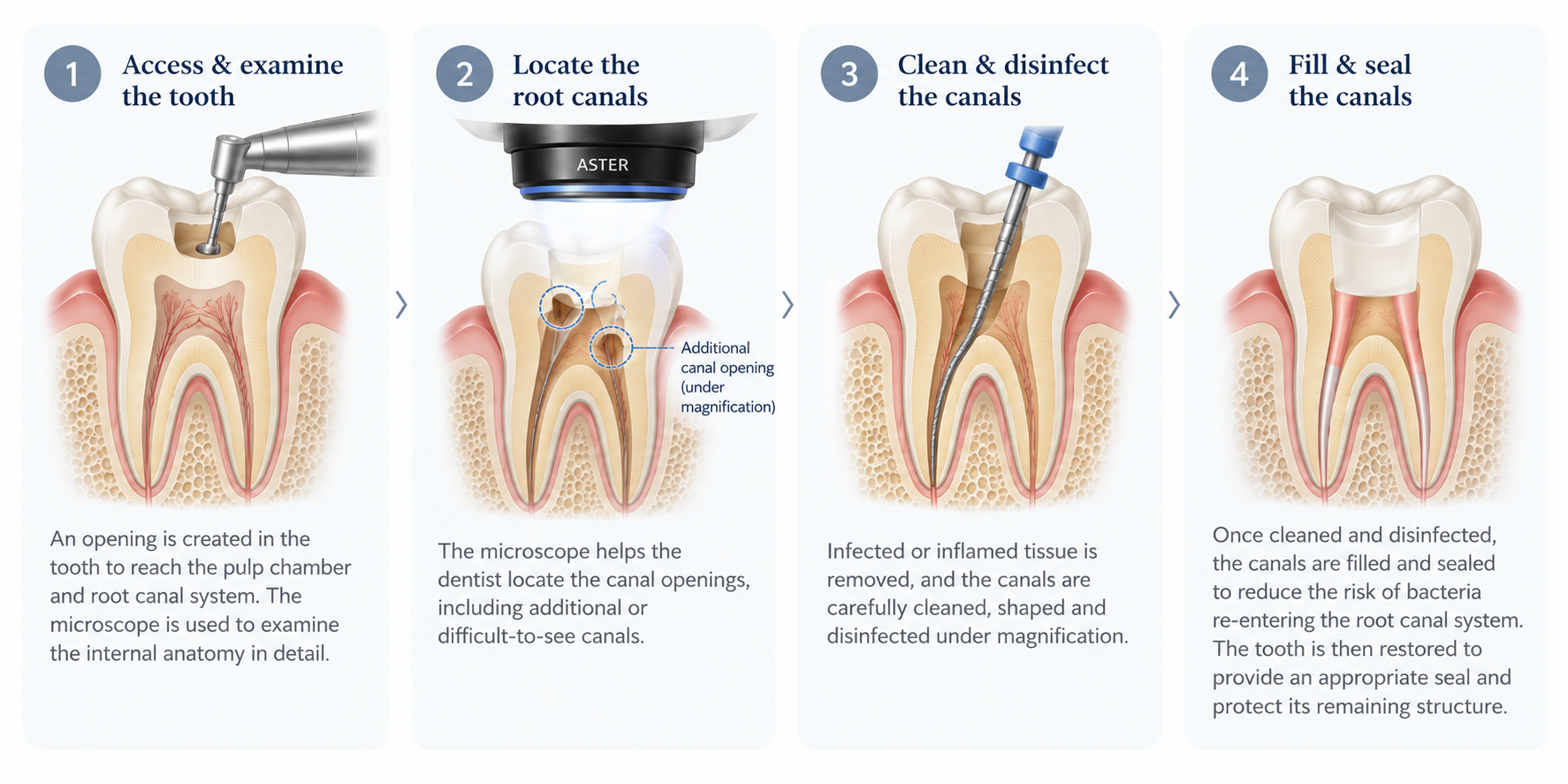
Microscope-assisted root canal treatment follows the same general principles as conventional root canal treatment, with a dental operating microscope used to provide enhanced magnification and illumination during key stages of the procedure.
Before treatment
The tooth is first assessed clinically and with dental imaging to understand the extent of infection or inflammation and the anatomy of the tooth. In more complex cases, additional imaging such as a CBCT scan can be used to provide a three-dimensional view of the roots and surrounding structures.
The treatment plan will be discussed with you before the procedure, including any findings that could affect the complexity of treatment.
During treatment
Local anaesthesia is administered to numb the tooth and surrounding area before treatment begins. Steps are taken throughout the procedure to keep you as comfortable as possible, and you can let the dental team know if you experience discomfort at any point.
The procedure then generally involves:
- Isolating the tooth — A rubber dam is placed around the tooth to keep the treatment area clean and prevent saliva and bacteria from entering the tooth during the procedure.
- Accessing and examining the tooth — A small opening is created to reach the pulp chamber. The dental operating microscope provides magnification and focused illumination as the internal anatomy is examined.
- Locating the root canals — The canal openings are identified under magnification, including any additional, narrow, calcified or difficult-to-see canals.
- Cleaning and disinfecting the canals — Infected or inflamed tissue is removed before the canals are carefully cleaned, shaped and disinfected.
- Examining the canals under magnification — The microscope allows the dentist to inspect the treatment area more closely for additional anatomy, debris or other features that require attention.
- Filling and sealing the canals — Once the canals have been adequately cleaned and disinfected, they are filled and sealed to reduce the risk of bacteria re-entering the root canal system.
After treatment
A temporary or permanent restoration is placed to seal the tooth after root canal treatment, depending on the treatment plan. Where a temporary restoration is used, the tooth will subsequently need to be permanently restored. Depending on the tooth and the amount of remaining structure, this can involve a filling, onlay or crown.
Some tenderness around the treated tooth can occur once the local anaesthetic wears off, particularly when biting or chewing. This generally improves as the tissues surrounding the tooth heal.
Depending on the condition of the tooth and whether infection was present around the root, follow-up can be recommended to assess healing.
Microscope-assisted vs conventional root canal treatment
The fundamental steps and goals of root canal treatment remain the same whether or not a dental operating microscope is used. The main difference is the level of magnification and illumination available to the dentist during treatment.
|
Factor |
Conventional root canal treatment |
Microscope-assisted root canal treatment |
|
Visualisation |
Treatment is performed using standard clinical lighting and visualisation, sometimes with other forms of magnification |
A dental operating microscope provides enhanced magnification and focused illumination |
|
Canal identification |
Root canals are identified using clinical examination, imaging and anatomical landmarks |
Magnification can help identify small, additional or difficult-to-see canal openings |
|
Complex anatomy |
Can be used to treat straightforward and some complex root canal systems |
Can provide additional visualisation when treating narrow, calcified or anatomically complex canals |
|
Retreatment |
Retreatment can be performed without a microscope depending on the case |
Particularly useful for locating missed canals and navigating changes or complications from previous treatment |
|
Precision |
Relies on the dentist’s clinical visibility and experience |
Enhanced visibility can support more precise treatment within the tooth |
|
Treatment goal |
Clean, disinfect and seal the root canal system |
Clean, disinfect and seal the root canal system |
Book a consultation
Advancements in dental technology have made it possible to perform root canal treatment with greater magnification and visualisation. At Aster Dental, microscope-assisted root canal treatment is available for selected root canal procedures and retreatment cases, particularly where the internal anatomy of the tooth is complex.
We understand that needing root canal treatment can feel daunting. Our dental team will explain what is happening with your tooth, discuss the treatment options available and take steps to keep you as comfortable as possible throughout your care.
Book a consultation with us to discuss your treatment options and the appropriate approach for your tooth.
Frequently asked questions
Is microscope-assisted root canal treatment different from a normal root canal?
The purpose and general steps of treatment are the same. The main difference is that a dental operating microscope provides enhanced magnification and illumination, allowing the dentist to see fine details within the tooth and root canal system more clearly during treatment.
Does every root canal need a microscope?
Not necessarily. Root canal treatment can be performed without a dental operating microscope. However, magnification can be particularly useful for teeth with complex anatomy, narrow or calcified canals, previous root canal treatment or other technical challenges.
Can a microscope help find missed root canals?
Yes. Magnification and focused illumination can help the dentist identify small or additional canal openings that can be difficult to see without enhanced visualisation. This can be particularly useful during root canal retreatment when a previously untreated canal is suspected.
Can a dental microscope help with calcified canals?
Yes. Calcification can make root canal openings narrow or difficult to locate. A dental operating microscope provides greater magnification and illumination, which can help the dentist identify and access these areas more precisely.
Can a dental microscope detect cracks in a tooth?
A dental operating microscope can help identify some cracks and structural defects that are visible within the accessible portions of the tooth. However, not all cracks can be detected using magnification alone, and other clinical findings or imaging can also be needed.
Is microscope-assisted root canal treatment more painful?
The use of a microscope itself does not make root canal treatment more painful. It is a visualisation tool used by the dentist during the procedure and does not change how local anaesthesia is administered.
How long does microscope-assisted root canal treatment take?
Treatment time varies depending on the tooth, the complexity of its root canal anatomy and whether the tooth has previously undergone root canal treatment. More complex cases, such as calcified canals or retreatment, can require additional time or more than one appointment.